<p style="text-align: justify;"><span style="font-family: Verdana; font-size: 11pt;">ELSEVIER SAUNDERS<br>THE DENTAL CLINICS OF NORTH AMERICA</span></p>
<h3>Low-level laser therapy in dentistry</h3>
<p align="center" style="text-align: center;"><span style="font-family: Verdana; font-size: 11pt;">Grace Sun, DDS, FAACD, FAGD</span></p>
<p class="MsoBodyText" style="text-align: justify;"> Therapeutic laser treatment, also referred to as low-level laser therapy (LLLT), offers numerous benefits. Along with the primary benefit of being non-surgical, it promotes tissue healing and reduces edema, inflammation, and pain. For more than 30 years, LLLT has been an interesting but not well-defined field among the medical, dental, physiotherapy, and veterinary professions.</p>
<p style="text-align: justify;"><span style="font-family: Verdana; font-size: 11pt;"> The lack of recognition among clinicians and researchers is due in part to the weakness of published materials. Some studies have ill-defined treatment parameters or poorly designed experiments with no control groups or double blind designs.</span></p>
<p class="MsoBodyText2"> Research needs to be done with the various wavelengths in creating different treatment effects and establishing a proper treatment protocol for each different situation. However, a growing number of clinicians use LLLT when treating patients because of the observed success. </p>
<p class="MsoBodyText2"> There are at least 2500 scientific studies in the field of light therapy. Among these are more than 100 positive, double blind studies. In 2001, the dental profession had conducted approximately 350 of these laser therapy studies from 98 institutions in 38 countries. Although the quality of these studies varies, more than 90% report positive effects of laser therapy.</p>
<h1>History and development</h1>
<p style="text-align: justify;"><span style="font-family: Verdana; font-size: 11pt;"> The roots of light therapy can be traced back to when our ancestors practiced heliotherapy, which gradually developed into actinotherapy and photomedicine (i.e. the use of UV radiation for sterilization). In 1903, Finsen was awarded the Nobel Prize for developing the carbon arc lamp incorporating lenses and filters for the treatment of lupus vulgaris.</span></p>
<p style="text-align: justify;"><span style="font-family: Verdana; font-size: 11pt;"> Subsequently, actinotherapy was used for treating tuberculosis and open wounds. In 1919, X-ray evidence led to the use of UV radiation in the treatment of rickets. Actinotherapy also stimulates the wound healing of ulcers, boils, and carbuncles and is used in the treatment of acne vulgaris and neonatal jaundice and for pain relief. Nevertheless, its potential carcinogenic side effects limit its usage.</span></p>
<p style="text-align: justify;"><span style="font-family: Verdana; font-size: 11pt;"> After Theodore Maiman in Los Angeles developed the first laser in 1960, there was a rapid development and interest in laser research. During the late 1960s in Budapest, Mester initiated studies on the possible carcinogenic effects of the low-power ruby and HeNe lasers. The mice he used in the study regrew hair faster than the controls, and the laser had no carcinogenic effect on the experimental group. Mester conducted a series of animal and laboratory studies. The most striking feature in the animal studies was that laser-irradiated experimental wounds healed better than non-irradiated wounds. Mester used this therapy for 875 patients with open wounds where conventional therapies had failed and had a success rate of 85%. Early experiments confirmed that the dosage follows the so-called Arndt-Schultz law: too small a dose gives no effect; there is a therapeutic window within a certain dose range; and doses over that range are inhibitory. Optimal doses for open wound were suggested to be 1 to 1 J/cm2. In 1973, Plog began to investigate the HeNe laser as a viable alternative to needles for acupuncture treatment. During the next decade, many research projects were conducted in Eastern Europe, the Soviet Union, and China.</span></p>
<p style="text-align: justify;"><span style="font-family: Verdana; font-size: 11pt;"> In the 1980s, clinical applications of the LLLT started to appear. Low-level lasers started gaining popularity in Europe, Asia, South America, and Australia, where the less-expensive and higher-powered output (30 mW or less) semiconductor (GaA1As or GaAs) devices were used. In the 1990s, increasingly powerful lasers at reasonable prices delivering higher doses proved to be more effective. The most recently FDA-approved treatment of carpal tunnel syndrome and "minor chronic neck and shoulder pain of musculoskeletal origin" has also proved to have a positive effect on the awareness and development of the therapeutic laser therapy.</span></p>
<h1>Terms and definitions</h1>
<p style="text-align: justify;"><span style="font-family: Verdana; font-size: 11pt;">The names to identify and differentiate therapeutic lasers from surgical lasers include soft, cold, low-intensity laser therapy, and LLLT. Therapeutic lasers are classified as class III medical devices, and surgical lasers are classified as class IV. Some phrases and phenomena describing the biologic effects of the therapeutic lasers are laser photobiostimulation, photo-biostimulation or biostimulation. In addition to the stimulating effect, the cellular effects also include bioinhibition, which can increase and decrease the physiologic functions to reach normalization. A more appropriate designation of the phenomenon might be laser photobiomodulation or laser bioactivation. The phrase "therapeutic laser" has also been used to suggest the purpose and intent of the treatment.</span></p>
<h1>Equipment</h1>
<p style="text-align: justify;"><span style="font-family: Verdana; font-size: 11pt;">LLLT benefits can be performed with various wavelengths and units with different outputs. Usually the therapeutic window for subthermal tissue interaction is 1 to 500 mW, but surgical lasers can be defocused and used as a "low level" laser.</span></p>
<p style="text-align: justify;"><span style="font-family: Verdana; font-size: 11pt;"> The first working laser, presented at a press conference arranged at the Hughes Aircraft Laboratory in Los Angeles on 7 July 1960, was a ruby laser (a solid-state laser using a single, rod-shaped ruby crystal). It emitted pulsed light at a wavelength of 694 nm. The ruby laser was also the first to be used in biostimulatory research in the mid 1960s. Among its successors was the HeNe (Helium-Neon) laser, a gas laser emitting at 632.8 nm with a power output of 1 to 5 mW. The HeNe laser was used predominantly in Eastern Europe and China in the mid to late 1970s.</span></p>
<p style="text-align: justify;"><span style="font-family: Verdana; font-size: 11pt;"> The most popular lasers are relatively inexpensive diode units that were developed in the 1980s. The GaAs (gallium-arsenide; 904 nm) diode laser was developed in the early 1980s and was typically 1 to 4 mW. Pulse-train modulated GaAs lasers entered the market in the late 1980s.</span></p>
<p style="text-align: justify;"><span style="font-family: Verdana; font-size: 11pt;"> The GaA1As (gallium-aluminum-arsenide; 780-890 nm) was developed in the late 1980s. It originally was designed as a 10- to 30-mW unit but since the late 1990s has been featured up to 500 mW.</span></p>
<p style="text-align: justify;"><span style="font-family: Verdana; font-size: 11pt;"> The InGaA1P (indium-gallium-aluminium-phosphide; 630-700 nm) diode lasers were developed in the mid 1990s. Typically 25 to 50 mW, they offer an alternative to the HeNe laser for surface wound healing.</span></p>
<p style="text-align: justify;"><span style="font-family: Verdana; font-size: 11pt;"> Combination probes of two laser wavelengths or one or more laser diodes with LEDs of various wavelengths may be made as "cluster probes." The effect of merging an incoherent LED with the laser requires further research to determine its effectiveness.</span></p>
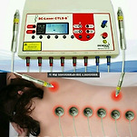
<p style="text-align: justify;"><span style="font-family: Verdana; font-size: 11pt;"> Dental therapeutic lasers are usually the size of an electric toothbrush and come with an attachable intraoral probe shaped like the wand used in composite curing light units (Fig. 1). The power of the GaA1As lasers should not be less than 100 mW to obtain the desired biologic effect in a reasonable time.</span></p>
<p style="text-align: center;"><br></p>
<h1>Dosage and calculations</h1>
<p style="text-align: justify;"><span style="font-family: Verdana; font-size: 11pt;"> Although there is a wide therapeutic window in laser therapy, it is essential to apply a reasonable dose. To calculate the dose (energy density), the given energy is calculated as mW x seconds (e.g., 100 mW x 10 seconds = 1000 mJ = 1 J). The dose is calculated by dividing the energy with the irradiated area. If this area is 1 cm<sup>2</sup>, the calculation is 1/1 = 1 J/cm2. If the irradiated area is 0.25 cm<sup>2</sup>, the calculation is 1 /0.25 = 4 J/cm2. A reasonable power density (mW per cm2) is necessary to trigger biologic effects, so low output cannot be fully compensated by longer exposure, and the depth of the treatment target site must be considered. Dose recommendations in seconds or minutes can be made only if the characteristics of specific laser are known. Laser light is reflected, absorbed, transmitted, and scattered after entering tissue. The amount of tissue between the laser probe and the target tissue and the type of tissue must be considered. For example, laser energy is more easily transmitted through mucosa and fat than through muscle. Hemoglobin and other pigments are strong absorbers of laser light and therefore require increased dosage. Penetration can be improved by using pressure, moving the laser closer to the target, or inducing a partial ischemia in the area. The skin coloration must also be considered because melanin is a strong absorber of light.</span></p>
<p style="text-align: justify;"><span style="font-family: Verdana; font-size: 11pt;"> Contact mode is needed for all applications with one exception. Treating an open wound requires a 2- to 4-mm separation distance between the laser and the target tissue. When contacting dental structures, some fluid might be needed to ensure full contact between the probe and the surface to minimize loss of energy.</span></p>
<p style="text-align: justify;"><span style="font-family: Verdana; font-size: 11pt;"> The following are some suggested treatment dosages: 2 to 3 J/cm2 two or three limes a week on gingival tissues; 4 to 6 J/cm2 two or three times a week on muscles; 6 to 10 J/cm2 once or twice a week on a TM joint; and 2 to 4 J/cm2 directly on the tooth or indirectly above the apex or osseous structure.</span></p>
<h1>The mechanisms</h1>
<p style="text-align: justify;"><span style="font-family: Verdana; font-size: 11pt;"> The principle of using LLLT is to supply direct biostimulative light energy to the body's cells. Cellular photoreceptors (e.g. cytochromophores and antenna pigments) can absorb low-level laser light and pass it on to mitochondria, which promptly produce the cell's fuel, ATP (Fig. 2).</span></p>
<p style="text-align: center;">
<br></p>
<p style="text-align: justify;"><span style="font-family: Verdana; font-size: 11pt;"> The most popularly described treatment benefit of LLLT is wound healing. From the studies of Mester et al, the electron microscope examination showed evidence of accumulated collagen fibrils and electron-dense vesicles intracytoplasmatically within the laser-stimulated fibroblasts as compared with untreated areas. Also, the measurement from the incorporation of 3H-thymidine showed accelerated cell reproduction and increased prostaglandin levels after irradiation. Increased microcirculation can be observed with the increased redness around the wound area; during the initial treatment stage, the patient can feel the transient pin-prickling sensation, which is thought to be evidence of the accelerated wound healing.</span></p>
<p style="text-align: justify;"><span style="font-family: Verdana; font-size: 11pt;"> The mechanisms of action underlying the analgesic effects remain unclear, despite the implicit treatment benefits. There is evidence suggesting that LLLT may have significant neuropharmacologic effects on the synthesis, release, and metabolism of a range of neurochemicals, including serotonin and acetylcholine at the central level and histamine and prostaglandin at the peripheral level. The pain influence has also been explained by the LLLT effect on enhanced synthesis of endorphin, decreased c-fiber activity, bradykinin, and altered pain threshold.</span></p>
<p style="text-align: justify;"><span style="font-family: Verdana; font-size: 11pt;"> Skepticism surrounds the analgesic effects due to conflicting results, obvious placebo potential, and dominating subjective findings. This is an important field for research and investigation.</span></p>
<p style="text-align: justify;"><span style="font-family: Verdana; font-size: 11pt;"> The most recognized theory to explain the effects and mechanisms of therapeutic lasers is the photochemical theory. According to this theory, the light is absorbed by certain molecules, followed by a cascade of biologic events. Suggested photoreceptors are the endogenous porphyrins and molecules in the respiratory chain, such as cytochrome c-oxidase, leading to increased ATP production.</span></p>
<p style="text-align: justify;"><span style="font-family: Verdana; font-size: 11pt;"> The photosensitivity of proteins is well known. There are more than 300 photochemically reactive proteins capable of harvesting low light energy. In humans, the most well known photochemically active receptor proteins are rod and cone pigments in the eye. However, other human photoreceptors, such as encephalopsin in the brain and pinopsin in the pineal gland, demonstrate the importance of light for human life.</span></p>
<p style="text-align: justify;"><span style="font-family: Verdana; font-size: 11pt;"> It is important to consider the following points when learning about the mechanisms of low-level laser therapy. First, the coherence of the electromagnetic energy plays a role in the efficacy of the treatment. The degree of coherence is related to the spectral narrowness of the light source. Although the spectral width of the gas HeNc laser is typically in focus, that of a laser diode is typically 1 to 10 nm, and the spectral width of LED is 30 to 100 nm. Noncoherent LED systems have a significant biologic effect. However, these effects seem to be limited to superficial tissues and, in some cases, to deeper tissues due to secondary effects through the release of metabolites. Polarization of the light has been shown to be beneficial in wound healing studies. The polarization of noncoherent light disappears shortly after entering tissue. The coherent character of the laser light is not lost after penetrating the tissue but is split into small coherent and polarized islands called speckles. The speckle pattern is maintained through the irradiated volume of tissue. Due to intensity differences within the speckle field, temperature and electric field gradients occur. Such gradients create a force on cells and organelles but do not affect the motion of atoms and molecules, as is the case in heat therapy. The gradient forces influence organelles such as mitochondria, which may enhance their interaction. This may explain why so many different lasers, including surgical lasers such as argon, Nd:YAG, diodes, and CO2 seem to have a stimulative/regulative effect on tissue that encompasses pain relief and wound healing. Properties such as penetration characteristics, light configuration, and power density are important factors. There are few in vivo studies comparing the effects of coherent and noncoherent light, but in all of them (approximately 20 studies) coherent light has come out on top.</span></p>
<p style="text-align: justify;"><span style="font-family: Verdana; font-size: 11pt;"> Surgical lasers typically have a central zone of carbonization, surrounded by zones of vaporization, coagulation, protein denaturation, and a stimulating zone. This may be one reason for the improved healing with laser surgery as compared with traditional scalpel surgery. Additionally, the effect of therapeutic laser irradiation is most prominent in cells in a reduced redox state. This means that the effect on healthy cells is less prominent and transient. Nevertheless, some investigators recommend irradiation before and after surgical interventions.</span></p>
<p style="text-align: justify;"><span style="font-family: Verdana; font-size: 11pt;"> Last, the effect of the laser is localized at the treatment site but can have a more generalized systemic effect. Infrared lasers have greater penetration than lasers in the visible range and are therefore able to affect deeper-lying conditions. However, the light does not need to reach the target cells to have the treatment effect. The CO2 laser has been used in the defocused mode as a biostimulator and has been used for deeper-lying conditions such as tendonitis. Because the light at that wavelength can penetrate skin less than 1 mm, the effect on deeper tissues is supposed to be influenced directly by blood metabolites. This addresses the systemic effect of the laser therapy. This theory postulates that a condition such as a burn treated on one hand influences a wound on the other hand but with less effect.</span></p>
<h1>Contraindications and safety measures</h1>
<p style="text-align: justify;"><span style="font-family: Verdana; font-size: 11pt;"> Therapeutic lasers have been used for more than 30 years. There are no reports of patients being harmed by therapeutic lasers, and class III lasers are termed nonsignificant risk medical devices. The risk of eye injury is minimal but must be considered, especially for high-output lasers in the invisible range. Diode laser light is generally divergent; however, if the light is collimated, the risk of eye injury increases significantly. Protective goggles, specific for the wavelength, must be used for the patient and the therapist.</span></p>
<p style="text-align: justify;"><span style="font-family: Verdana; font-size: 11pt;"> Although there are no contraindications reported for dental therapeutic lasers, some caveats and side effects exist. Suspected malignancies should never be treated by anyone but the specialist. Because laser light affects several rheologic factors, patients with coagulation disorders need special attention. Patients with chronic pain have reported increased tiredness for a brief period, and long-standing pain conditions may transiently increase.</span></p>
<h1> Clinical applications</h1>
<p style="text-align: justify;"><span style="font-family: Verdana; font-size: 11pt;"> By understanding the basic cellular effects of the lasers and the intended treatment goal of reducing inflammation, accelerating the healing process, and providing pain relief, the general principles of application for various clinical conditions becomes clear. Beneficial treatment effects have been applied in dermatologic conditions such as wounds and inflammations, neural ailments in various locations, and musculoskeletal ailments causing pain and degeneration in various sites. Veterinary uses of LLLT include treating dogs and horses with nerve injuries, tendonitis, arthritis, and trauma from training and competition.</span></p>
<p style="text-align: justify;"><span style="font-family: Verdana; font-size: 11pt;"> The application of LLLT in dentistry is included with various clinical conditions. The general rule for intra-oral treatments is to use 2 to 4 J with the intra-oral probe (Fig. 3) and 4 to 10 J for extra-oral treatments</span></p>
<table width="100%" bordercolor="#111111" style="border-collapse: collapse;" border="0" cellspacing="0" cellpadding="0">
<tbody>
<tr>
<td width="50%" valign="top">
<p align="center"><br></p></td>
<td width="50%" valign="top">
<p align="center"><br>
<p align="center"> </p></td></tr></tbody></table>
<h1><i>Postoperative therapy and care</i></h1>
<p style="text-align: justify;"><span style="font-family: Verdana; font-size: 11pt;"> The purpose of using LLLT as part of postoperative therapy is to provide patients with the highest quality of health care. This should include minimal discomfort or pain and a shortened healing period. It can be applied to many dental procedures, such as operative dentistry, fixed prosthetics, nonsurgical and surgical endodontic procedures, nonsurgical and surgical periodontal treatments, implantology, oral surgery, or orthodontic adjustments.</span></p>
<p style="text-align: justify;"><span style="font-family: Verdana; font-size: 11pt;"> Approximately 2 J of energy are applied from the wand-like probe to the patient's injection and operative sites, apical to the tooth apex buccally and lingually, around the CEJ and masticatory muscles (Fig. 5). These can be immediate postoperative applications or continuing once or twice weekly treatments if the patient has had more extensive surgical procedures or for implant cases. For orthodontic adjustments, the treatment would be for every adjustment visit.</span></p>
<p style="text-align: center;"><br></p>
<h2> Reported effects of low-level laser therapy </h2>
<p style="text-align: justify;"><span style="font-family: Verdana; font-size: 11pt;"> <i>Temporomandibular disorders.</i> Diagnosis is essential before treatment is rendered. The infrared laser is effective for reducing pain and tension in the masticatory muscles, especially in cases of trismus. Symptomatic tender points and muscle attachments are treated with 6 to 10 J. After treatment, an increase in the range of movement has been noted. An interesting application is the treatment of somatosensory tinnitus, a condition where TMD therapy can play a vital role. The lateral pterygoid muscle is typically affected in these cases; laser irradiation of this and other involved muscles can lead to a rapid reduction of tension and pain in the muscular system. As co-intervention in arthralgic cases, lasers can be used to advantage.</span></p>
<p style="text-align: justify;"><span style="font-family: Verdana; font-size: 11pt;"> <i>Hypersensitive dentin</i>. Although many desensitizing agents treat this condition, the laser can treat more difficult cases. Kimura has published a review of the literature. A hypersensitive tooth that does not respond to 4 to 6 J per root in two or three sessions is indicated for endodontic treatment. The occlusal scheme should be eval‎uated as part of the treatment protocol.</span></p>
<h1><i>Postextraction and bone-healing therapy</i></h1>
<p style="text-align: justify;"><span style="font-family: Verdana; font-size: 11pt;"> It is useful to irradiate the area before and after an extraction. Irradiating before the extraction with 1 J at the injection site and 2 J right below the apices induces a transient but useful effect (i.e., a faster onset of local analgesia and reduced bleeding during and post extraction). After extraction, an additional 2 J cm2 on the alveolar and gingival tissues is needed to control the swelling and inflammation. Less postoperative pain and better healing can be expected.</span></p>
<p style="text-align: justify;"><span style="font-family: Verdana; font-size: 11pt;"> Ohshiro has conducted clinical and laboratory studies with different LLLT wavelengths on the activation of alveolar bone production. Nd:YAG, HeNe, and GaA1As diode have shown faster wound healing and bone formation after tooth extraction compared with unlased cases. HeNe and diode lasers showed similar or better effects than the Nd:YAG instrument. Experiments with Donryu rats demonstrated that LLLT with Nd:YAG, HeNe, and GaA1As diode devices produced nonthermal bioactive reaction in the irradiated bone defect and underlying marrow. This resulted in earlier osteogenesis than the control and bone treated with the CO2 laser. The excimer laser induced necrosis and delayed healing. The 248-nm beam can ionize living tissue. Another experiment on the LLLT effects on bone repair activation in laboratory mice used a bone inductive substance, bone morphogenetic protein (BMP). It concluded that in addition to the stimulating action of the BMP. LLLT stimulates undifferentiated mesenchymal cells into osteoblasts, resulting in increased osteogenesis. The increased blood circulation after LLLT might also bring a better supply of inorganic salts, promoting better bone formation.</span></p>
<h2>Orthodontics</h2>
<p style="text-align: justify;"><span style="font-family: Verdana; font-size: 11pt;"> In addition to reducing the initial tension pain, laser therapy can increase the speed of tooth movements by increased osteoclastic activity on the pressure side and increased osteoblastic activity on the tension side. Laser therapy has also been used for oral ulcerations induced by fixed orthodontic appliances.</span></p>
<h2>Herpes labialis</h2>
<p style="text-align: justify;"><span style="font-family: Verdana; font-size: 11pt;"> Herpes simplex virus infection of the lips is most common among adolescents and adults. Symptoms can range from mild discomfort to extreme pain. Soft lasers have been shown to have an effect similar to Acyclovir and have been shown to be effective if given in the silent periods between attacks. If applied in the prodromal stage, the blister is likely to disappear in 2 to 3 days with little discomfort, rather than 8 to 14 days. LLLT also reduces the frequency of recurrence and relapse rate. Zosters and postherpetic neuralgia may also be treated.</span></p>
<h2> Aphthous ulcers</h2>
<p style="text-align: justify;"><span style="font-family: Verdana; font-size: 11pt;"> LLLT for the treatment of aphthae can be recommended for its pain-relieving effect and the shortened healing time. The treatment dosage is the same for herpes and aphthae: 2 J/cm2 applied near contact. Repeat the following day.</span></p>
<p style="text-align: justify;"><b><i><span style="font-family: Verdana; font-size: 11pt;">Mucositis</span></i></b></p>
<p style="text-align: justify;"><span style="font-family: Verdana; font-size: 11pt;"> Patients undergoing radiation therapy or chemotherapy develop mucositis. Mucositis is painful and may force the oncologist to reduce the dosage or number of sessions. Red laser light has been shown to reduce the severity of mucositis and can be used prophylactically before radiation. The laser can also treat dermatitis induced by radiation therapy.</span></p>
<p style="text-align: justify;"><b><i><span style="font-family: Verdana; font-size: 11pt;">Paresthesia</span></i></b></p>
<p style="text-align: justify;"><span style="font-family: Verdana; font-size: 11pt;"> One of the risks of dental surgery is nerve injury and paresthesia, especially of the inferior alveolar nerve. Although most of these complications are transient, some are long standing or permanent. Using LLLT when the paresthesia has occurred, and prophylactically after surgery in a risk-involved zone could reduce these problems.</span></p>
<h2>Trigeminal neuralgia</h2>
<p style="text-align: justify;"><span style="font-family: Verdana; font-size: 11pt;"> LLLT has been documented to have a pain-relieving effect on trigeminal neuralgia (Fig. 6). In a double-blind study, 16 patients were treated weekly for 5 weeks. After that period 10 patients were free from pain, and two had noticeably less pain; four patients were unchanged. At one-year follow-up, six patients were free from pain</span><br></p>
<p style="text-align: justify;"><b><i><span style="font-family: Verdana; font-size: 11pt;">Nausea</span></i></b></p>
<p style="text-align: justify;"><span style="font-family: Verdana; font-size: 11pt;"> Some clinicians practice acupuncture. Although lasers can be used in the place of needles, only trained individuals should perform acupuncture. There is, however, a useful and risk-free point on the wrist, known by acupuncturists as the meridian point P 6 (Fig. 7). Three to four Joules on this point reduces gagging reflexes in most patients.</span></p>
<p style="text-align: center;">
<br></p>
<h2>Periodontitis</h2>
<p style="text-align: justify;"><span style="font-family: Verdana; font-size: 11pt;"> The use of LLLT helps to control the symptoms and condition of periodontitis. The anti-inflammatory effect slows or stops the deterioration of periodontal tissues and reduces the swelling to facilitate the hygiene in conjunction with other scaling, root planning, curettage, or surgical treatment. As a result, there is an accelerated healing and less post-op discomfort. Studies report stimulation of human periodontal fibroblasts, reduced gingivitis index, pocket depth, plaque index, gingival fluid, and metalloproteinase-8 levels (Qadri T, Miranda L. Tuner J, Gustafsson A. unpublished data. 2004), as well as positive results after gingivectomies. In the latter study, biometrical eval‎uation showed improvement of healing for the period of 21 and 28 days in the lased group. Clinical eval‎uation showed better reparation mainly after the third day for the active group.</span></p>
<h2>Nonbiostimulative effects</h2>
<p style="text-align: justify;"><span style="font-family: Verdana; font-size: 11pt;"> In addition to their ability to treat pain, edema, and inflammation, these lasers enhance the fluoride release from cements and varnishes and kill bacteria in the presence of suitable photosensitizing agents or various dyes. The latter is an example of photodynamic therapy, a longtime-practiced therapy in oncology.</span></p>
<h1>Summary</h1>
<p style="text-align: justify;"><span style="font-family: Verdana; font-size: 11pt;"> This introduction to LLLT briefly discusses more than three decades of international experimental and clinical research. No true side effects of using the low-level laser light have been found. When low-level laser light provides the energy that interacts with our cells, it creates a myriad of positive functions, such as accelerated wound healing, pain relief, regeneration, and immune enhancement. It is noninvasive, nonpharmaceutical, and economical. These benefits may help generate interest among more clinicians, researchers, and manufacturers to study and gain more knowledge on how best to use this phenomenon. Developing the equipment and treatment protocols and training the general educators and health practitioners is essential for improving health services and treatment outcomes.</span></p>
<!-- -->